A 10-year-old intact female Border Collie dog was presented for evaluation of abdominal distension. The dog had pale mucous membranes, and tachypnea.
A 10-year-old intact female Border Collie dog was presented for evaluation of abdominal distension. The dog had pale mucous membranes, and tachypnea.
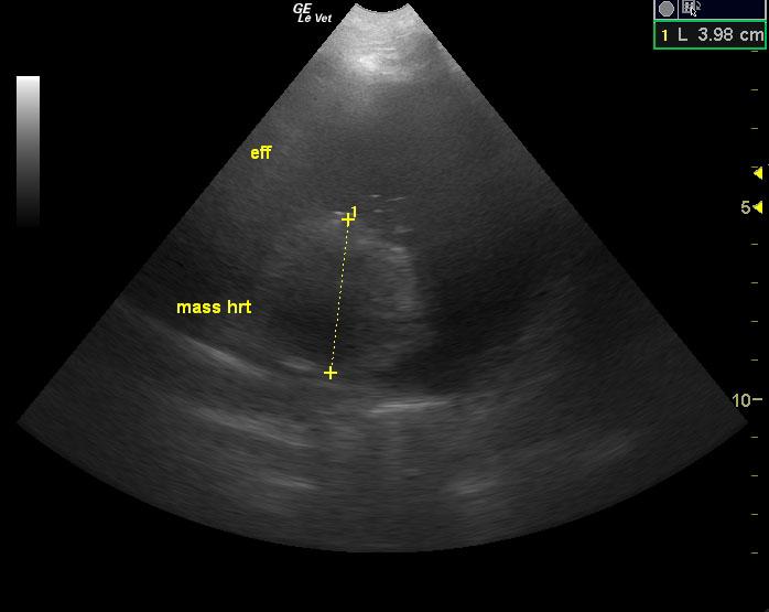
An aortic base tumor is likely given the location and the patient’s breed. Right-sided tamponade was noted. There is pericardial effusion and a valvular thrombus in RVOT.
The LV was normal in diastole and systole. The fractional shortening (FS) was within normal limits indicating adequate systolic function. The EPSS (end point to septal separation) was also within normal limits (another indicator of adequate systolic function). The endocardial surface of all chambers was normal in echogenicity and smooth. The MV showed normal excursion and anatomy. There was no significant mitral insufficiency noted on Doppler. The LA was normal in size. The La/Ao ratio was within normal limits for the patient’s body weight. The RV demonstrated normal thickness when compared to the LV (approx 1/3 of LV). There was a thrombus visible in the right ventricular outflow tract. The left ventricular outflow tract was normal in appearance. The MPA was normal in relation to the aorta (1:1) PA/Ao ratio). There were no heartworms noted in the visible portion of the pulmonic outflow tract. It had normal laminar patterns showing no evidence of stenosis or regurgitation. The PV was visualized and was normal. The myocardium throughout the heart was uniformly echogenic without evidence of significant fibrosis or infiltrative disease. There was no evidence of heart base or mediastinal masses in the visible ultrasonographic window. There was marked pericardial effusion present with a mass effect that was nearly 4 cm in diameter.
Splenic disease: neoplasia, torsion. Pericardial effusion. Peritonitis.
50 ml of pericardial effusion was drained from the pericardial sac. 12 ml of clear, transudative fluid was drained from the abdomen. No evaluations were performed on fluid.