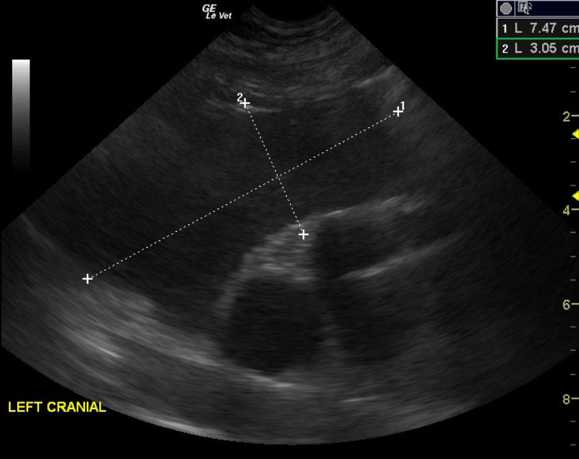
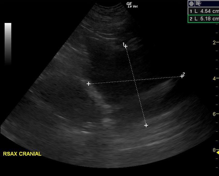
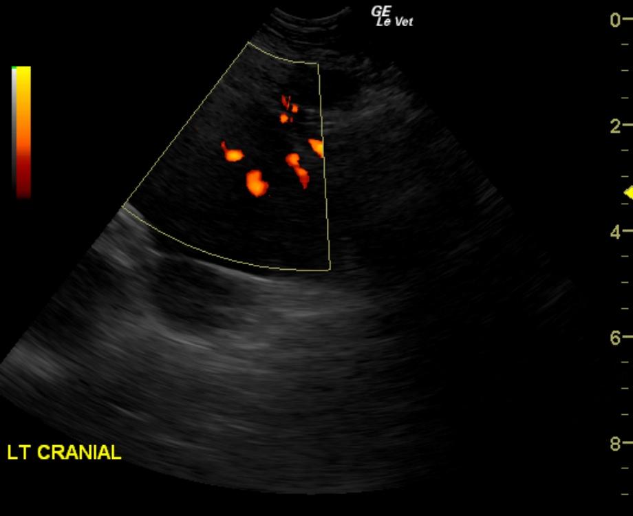
The patient is a Labrador Cross, SF, 12 years old. Concern for possible mediastinal mass per radiographs, limping on right front leg.
Sonographic Differential Diagnosis
Cranial mediastinal masses, suspect lymphoma, possible Thymoma. FNA were performed without complication.
Possible lung mass; however, no significant peripheral air entrapment was noted. Therefore lymph node origin is suspected. Recommend oncology consultation and abdominal screening for primary neoplasia as this may represent a metastatic pattern through the lymphatic tract.
Image Interpretation
Cranial mediastinal lymph node mass was noted and measured 7.4 x 3.0 cm on the left cranial and another mass in the right cranial region was noted and measured 5.2 x 4.5 cm. This is strongly suggestive for lymphoma.