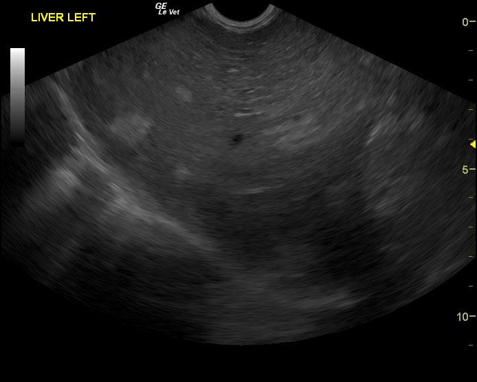
A 13-year-old FS Springer Spaniel that had been vomiting intermittently after a dental and skin mass removal that had been done a few days previously presented for an examination. The only abnormality on physical examination was a slight pot bellied appearance. Urinalysis showed an inappropriate specific gravity (1.018), bilirubinurea, proteinurea, and bacteriuria. CBC and serum biochemistry showed thrombocytosis, elevated ALP, elevated GGT, and elevated ALT activity, elevated cholesterol and elevated triglycerides. T4 was normal.
A 13-year-old FS Springer Spaniel that had been vomiting intermittently after a dental and skin mass removal that had been done a few days previously presented for an examination. The only abnormality on physical examination was a slight pot bellied appearance. Urinalysis showed an inappropriate specific gravity (1.018), bilirubinurea, proteinurea, and bacteriuria. CBC and serum biochemistry showed thrombocytosis, elevated ALP, elevated GGT, and elevated ALT activity, elevated cholesterol and elevated triglycerides. T4 was normal.