A 9-year-old SF Chihuahua was presented for————. Additional history was that the patient is fed on a vegan diet. CBC was within normal limits, whereas serum biochemistry showed mild elevation of ALP activity and hypertriglyceridemia.
A 9-year-old SF Chihuahua was presented for————. Additional history was that the patient is fed on a vegan diet. CBC was within normal limits, whereas serum biochemistry showed mild elevation of ALP activity and hypertriglyceridemia.
Case Study
06-00066 Milla C Cystitis and calculi—NEEDS HX, CDX—–
Sonographic Differential Diagnosis
Bladder calculus. Chronic cystitis pattern. Mild to moderate degenerative renal changes with mineralization. Left kidney medullary calculi. Vacuolar hepatopathy of the liver with minor remodeling.
Image Interpretation
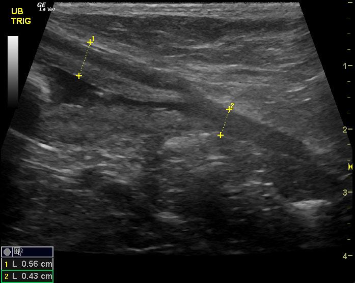
The urinary bladder in this patient revealed a shadowing 1.0 calculus and a chronic cystitis pattern was noted in the bladder wall that measured 0.62 cm with areas of echogenic remodeling in the mucosa. This is consistent with fibrosis. Cystotomy, stone analysis, body wall and culture would all be warranted with a minimal possibility of transitional cell carcinoma in the wall itself. However, these changes are most consistent with chronic cystitis. Long term antibiotic therapy would e necessary in this patient. Stone culture or bladder wall culture would be warranted. The pelvic urethra was mildly thickened in this patient owing to chronic irritation. The kidneys revealed largely normal size and structure, corticomedullary definition and ratio (cortex 1/3 of medulla) were essentially maintained with some age related loss of curvilinear pattern. The cortices presented largely uniform texture with some age related echogenic changes that are not likely of clinical significance at this time. Medullary echogenicity differed distinctly from that of the cortex and no evidence or dilation could be seen. The capsules were acceptably uniform for this age patient without dramatic irregularities. The left kidney measured 3.8 cm. Medullary calculi were noted in the left kidney. The right kidney measured 3.8 cm.
DX
Bladder calculus. Chronic cystitis pattern. Left kidney medullary calculi.
Outcome
Recommend cystotomy, bladder wall or stone analysis and bladder wall biopsy. The patient had a cystotomy and the stones were revealed to be struvites.
Sampling
The patient had a cystotomy and the stones were revealed to be struvites.


