A 5-year-old MN DSH cat was presented at an emergency facility for acute onset respiratory distress, vomiting, and lethargy. Abnormalities on physical examination were dyspnea with a respiratory rate of 80, muffled heart sounds, marked bilateral pulmonary crackles and wheezes, marked abdominal breathing, and red-tinged fluid in both nares. CBC and blood chemistry were within normal limits. PCV/TP was 48/6.0.
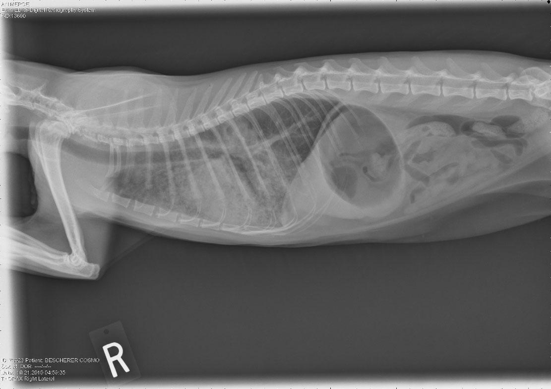
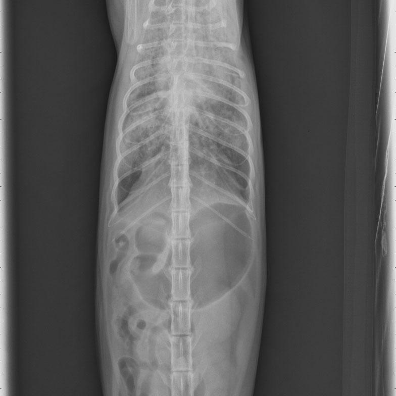
A 5-year-old MN DSH cat was presented at an emergency facility for acute onset respiratory distress, vomiting, and lethargy. Abnormalities on physical examination were dyspnea with a respiratory rate of 80, muffled heart sounds, marked bilateral pulmonary crackles and wheezes, marked abdominal breathing, and red-tinged fluid in both nares. CBC and blood chemistry were within normal limits. PCV/TP was 48/6.0. Survey radiographs showed border effacement of the cardiac silhouette, bilateral patchy pulmonary infiltrates, aerophagia, and a moderate amount of gas dispersed throughout the small intestine.
Case Study
Lung consolidation and mixed inflammation in a 5 year old MN DSH dyspneic cat
Sonographic Differential Diagnosis
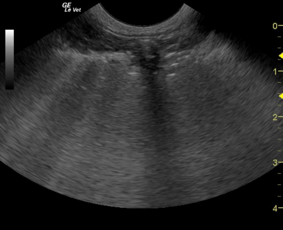
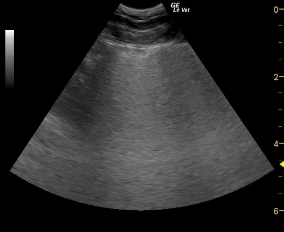
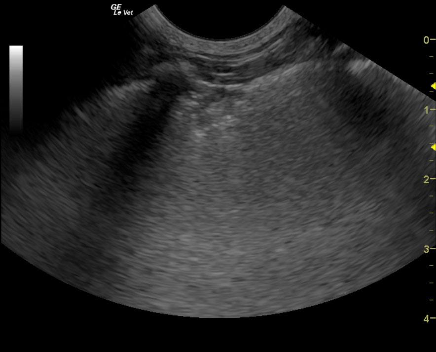
Undefined “shower curtain” lung pattern with multi-focal consolidation.
Image Interpretation
The cardiac presentation in this patient was essentially normal. Investigation of primary lung disease is of concern, which is why fine-needle aspirates were performed. “Shower curtain” lung pattern was noted in this patient. This is consistent with pulmonary edema, possible pneumonitis, metastatic disease or fungal disease. Other causes of pneumonitis should be investigated. 25-gauge aspirates were performed and submitted for cytology review.
DX
Outcome
It was recommended that the patient be stabilized on bronchiodilators, broad spectrum antibiotics, and aspirin pending cytologic results. A recheck echocardiogram was also recommended within 48-72 hours. The patient was treated with Lasix, Zithromax, Ampicillin, Albuterol, Clopidogrel, Flovent, and nebulization with Gentocin. After the patient was stabilized and breathing well outside of oxygen therapy, he was discharged with Lasix, Enacard, Zithromax, Clopidogrel, and instructions to follow-up with the referring veterinarian.
Clinical Differential Diagnosis
Acute lung edema – heart disease (DCM/HCM/myocarditis), smoke/chemical inhalation, trauma, toxins, fluid overload, anaphylaxis, ARDS, neoplasia Acute pneumonia – bacterial, viral, aspiration, inhalation
Sampling
US-guided FNAs of the consolidated lung tissue revealed mixed inflammation with mesenchymal cell proliferation.
Video
Patient Information
Clinical Signs
- Lethargy
- Tachypnea
- Vomiting
Exam Finding
- Abnormal lung sounds
- Pulmonary crackles
- Respiratory Distress
- Tachypnea
Images
Clinical Signs
- Lethargy
- Tachypnea
- Vomiting