An 11-year-old intact female Cairn terrier dog was presented on emergency at RDVM for vomiting bile for 24 hours. Abnormalities on physical examination were lethargy, severe dental disease, prominent popliteal lymph nodes, and a tense abdomen and prominent liver on palpation. CBC and blood chemistry showed marked leukocytosis, neutrophilia, monocytosis, eosinopenia, thrombocytosis, elevated total proteins, hyperglobulinemia, markedly elevated ALP activity, hyperbilirubinemia, and mild hypercholesterolemia. On abdominal radiographs gas in the stomach and hepatomegaly were evident.
An 11-year-old intact female Cairn terrier dog was presented on emergency at RDVM for vomiting bile for 24 hours. Abnormalities on physical examination were lethargy, severe dental disease, prominent popliteal lymph nodes, and a tense abdomen and prominent liver on palpation. CBC and blood chemistry showed marked leukocytosis, neutrophilia, monocytosis, eosinopenia, thrombocytosis, elevated total proteins, hyperglobulinemia, markedly elevated ALP activity, hyperbilirubinemia, and mild hypercholesterolemia. On abdominal radiographs gas in the stomach and hepatomegaly were evident. The patient was treated with IV fluids, Ampicillin, Baytril, Famotadine, Cerenia, and Buprenex.
Case Study
03_00222 Lucy P Mucocele, cholangiohepatitis
Sonographic Differential Diagnosis
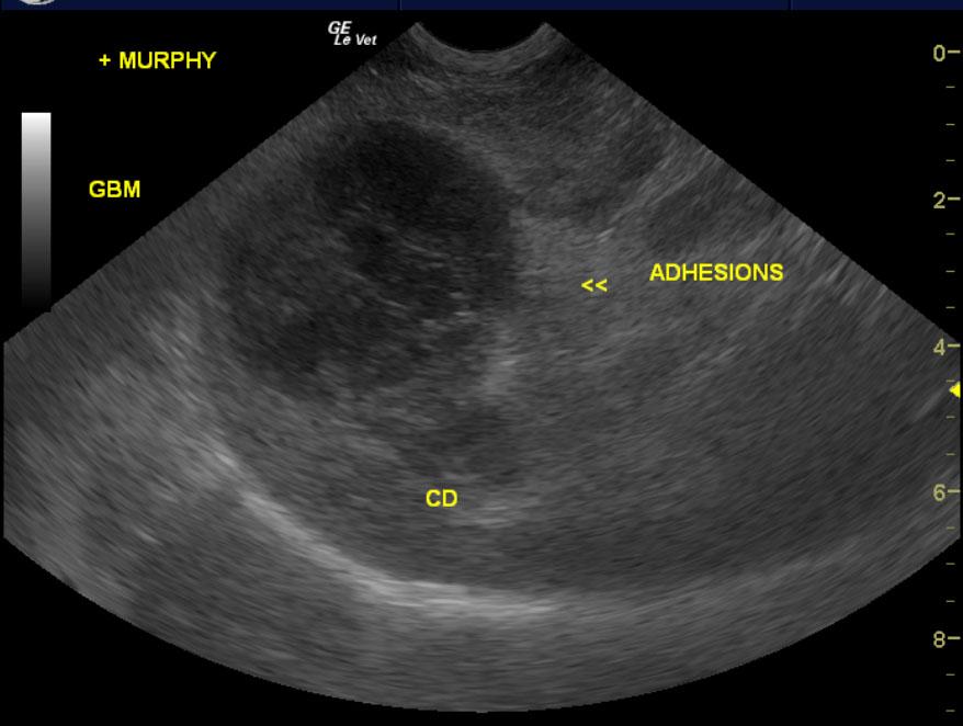
Inflamed gallbladder mucocele with dilated common bile duct and cholangiohepatitis pattern.
Image Interpretation
The liver was swollen in contour with a 3.0 cm region around the gallbladder and common bile duct where there was ill-defined omentum as noted. This is consistent with adhesions. The liver presented mild heterogenous changes and should be biopsied at the time of surgery. Chronic inflammatory hepatopathy is suspected. The gallbladder was severely dilated with dilated cystic duct. This is consistent with inflammation and potential history of perforation and gallbladder mucocele.
DX
Outcome
The patient was recommended for cholecystectomy, possible common bile duct reconstruction, and cultures and biopsy of the gallbladder and liver. The patient underwent an exploratory surgery with cholecystectomy, liver biopsy, ovariohysterectomy, and MGT removal. Other than needing to be syringe fed post-operatively, the patient was doing very well. At sutures out appointment a few weeks later, the owner reported the patient as doing great. Physical examination found the patient BAR with the incision healed well. Abnormalities on follow-up blood work were mild neutrophilia, ongoing thrombocytosis, and markedly elevated ALP and GGT activity. The patient was discharged with Amoxicillin, Metronidazole, and Denamarin.
Clinical Differential Diagnosis
Liver – infectious (bacteria/viral/fungal), neoplasia, toxin, Cushing’s disease, trauma Gall bladder – mucocele, cholicystitis, stone, bile duct obstruction Duodenal disease – foreign body, neoplasia, duodenitis, ulceration
Sampling
Full-thickness surgical biopsies of the gall bladder revealed purulent pericholecystitis, liver sample revealed marked, purulent, subacute cholangiohepatitis, and the mammary mass was simple mammary carcinoma.
Video
Patient Information
Clinical Signs
- Lethargy
- Vomiting
Exam Finding
- Abdominal Distension
- Enlarged Lymph Nodes
- Hepatomegaly
- Lethargy
- Tense Abdomen
Images
Blood Chemistry
- Alkaline Phosphatase (SAP), High
- Globulin, High
- Total Protein, High
CBC
- Eosinophils, Low
- Monocytes, High
- Neutrophils, High
- Platelet Count, High
- WBC, High
Clinical Signs
- Lethargy
- Vomiting