This 11-year-old FS Springer Spaniel was presented for straining to urinate and abdominal distension. No other symptoms were noted by the owner. The physical exam revealed a large palpable abdominal mass. Radiographs revealed a large soft tissue mass but the origin was not evident. Blood analysis demonstrated mild anemia and mild leukocytosis with a left shift.
This 11-year-old FS Springer Spaniel was presented for straining to urinate and abdominal distension. No other symptoms were noted by the owner. The physical exam revealed a large palpable abdominal mass. Radiographs revealed a large soft tissue mass but the origin was not evident. Blood analysis demonstrated mild anemia and mild leukocytosis with a left shift.
Case Study
Intestinal Stromal Tumor in an 11 year old FS Springer Spaniel dog
Sonographic Differential Diagnosis
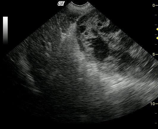
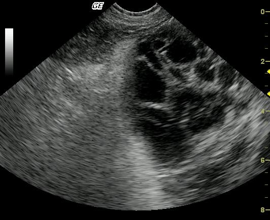
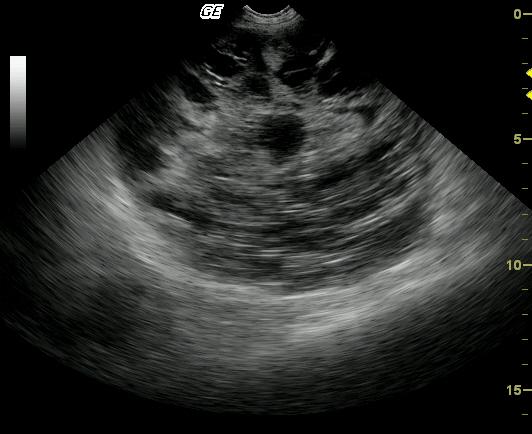
Large cavitated mass with bordering inflammation and suspect omental and mesenteric adhesions. Neoplasia, either benign or malignant arising from a regional soft tissue structure, mesentery or omentum is primarily suspected. Also considered are inflammatory, infectious (parasitic?), granulomatous, hematoma.
Image Interpretation
A large, well-encapsulated, cavitated and septated mass is present within the peritoneal cavity. The mass is bordered by echogenic omentum and mesentery, which may be adhered. The origin of the mass is not apparent on these images.
DX
Intestinal stromal tumor
Outcome
The origin of the mass could not be discerned by the sonogram even though it enveloped intestine and mesentery. The patient is clinically normal at a 5-month follow-up. A repeat sonogram was suggested but had not been performed. After surgical resection, no adjunctive therapy was administered.
Comments
No video is available on this patient.
Clinical Differential Diagnosis
Neoplasia, pancreatic sequestrum, lipoma.
Sampling
Surgical exploratory was performed after the sonogram defined a lack of evident metastatic disease to any organ. The mass was found to be of intestinal origin and was fully resected.
Patient Information
Patient Name :
Frannie H
Gender :
Female, Spayed
Species :
Canine
Type of Imaging : Ultrasound
Status :
Complete
Liz Wuz Here :
Yes
Code :
04_00094
Clinical Signs
- Abdominal Distension
- Stranguria
Exam Finding
- Abdominal Distension
- Palpable mass
Images
CBC
- Left Shift
- RBC, Low
- WBC, High
Clinical Signs
- Abdominal Distension
- Stranguria